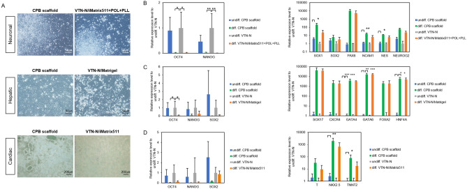
Figure 8.
Differentiation of human induced pluripotent stem cells on CPB scaffold into three-germ layers. (A) Representative images of differentiated 253G1 cells at 6–7 days after neuronal ectoderm (top), hepatic endoderm (middle), and cardiac mesoderm (bottom) induction. Left images show the differentiated cells on CPB scaffold induced from the cells maintained on CPB scaffold more than 10 passages before differentiation induction. Right images show differentiated cells on control differentiation substrate according to each protocol from the cells maintained on the control maintenance substrate VTN-N. The control differentiation substrate was Laminin/poly-L-ornithine/poly-L-lysin (iMatrix511/POL/PLL) for neural, Matrigel for hepatic, and laminin (iMatrix-511) for cardiac differentiation. Scale bars are indicated in the images. (B) Expression level of undifferentiated (OCT4 and NANOG) and neural lineage (SOX1, SOX2, PAX6, NCAM1, NES, and NEUROG2) marker genes in the neuro-ectodermal differentiation. (C) Expression level of undifferentiated (OCT4, NANOG, and SOX2) and hepatic lineage (SOX17, CXCR4, GATA4, GATA6m FOXA2, and HNF4A) maker genes in the hepatic endoderm differentiation. (D) Expression level of undifferentiated (OCT4, NANOG, and SOX2) and cardiac lineage (T, NKX2.5, and TNNT2) marker genes in the cardiac mesoderm differentiation. All gene expression levels indicated were examined by RT-qPCR, and indicated as relative expression level to undifferentiated 253G1 cells cultured on VTN-N. Data are presented as mean ± SD of three independent experiments. (*P < 0.05, **P < 0.01 and ***P < 0.001 compared to undifferentiated 253G1 cells cultured on VTN-N).

