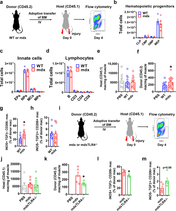
Fig. 7. Adoptive transfer reveals the altered phenotype of mdx and mdxTLR4–/– BMDM after muscle injury in vivo.
a Pictorial representation showing adoptive transfer of bone marrow (BM) from WT or mdx (both CD45.2 allele) mice into non-irradiated congenic WT (CD45.1 allele) mice at the onset of cardiotoxin-induced hindlimb muscle injury, followed by flow cytometric analysis of donor and host BMDM in the injured muscle 4 days later. b–d Comparison of absolute number of: b Hematopoietic precursor cells (CLP: common lymphoid progenitors, CMP: common myeloid progenitors, GMP: granulocyte/macrophage progenitors, MEP: megakaryocyte/erythrocyte progenitors; n = 5/group), c Innate myeloid cells (EPs: Eosinophils, NPs: Neutrophils, MCs: Monocytes, MPs: Macrophages) (n = 5/group) and d Lymphocytes (B: B cells, CD3: CD3 T cells, CD4: CD4 T cells, CD8: CD8 T cells) (n = 5/group) in the BM of age-matched WT or mdx at necrotic phase. e, f Quantification of host (CD45.1–WT; e n = 15/group) and donor (CD45.2-either WT or mdx or mock adoptive transfer with PBS; f n = 14 for WT, rest n = 15/group) macrophages in the injured muscles (defined as CD45 + CD11c-CD11b + F4/80 + ). g Percentage of donor pro-inflammatory (iNOS + TGFβ-CD206-) and h donor anti-inflammatory (iNOS-TGFβ + CD206 + ) BMDM of either WT or mdx origin in the injured WT host muscle (n = 10/group). i Schematic showing adoptive transfer of mdx or mdxTLR4–/– BM using the same experimental design. j, k Quantification of host (CD45.1–WT; n = 6 for PBS, rest n = 10/group) and donor (CD45.2-either mdx or mdxTLR4–/–; n = 5/group) macrophages in the injured muscles. l Percentage of donor pro-inflammatory and m donor anti-inflammatory BMDM of either mdx or mdxTLR4-/- origin in the injured WT host muscle (n = 5/group). Data represent means ± SEM of biologically independent samples from different mice. *P < 0.05 (unpaired t-test, two-tailed). See Source Data file for the exact P-values.

