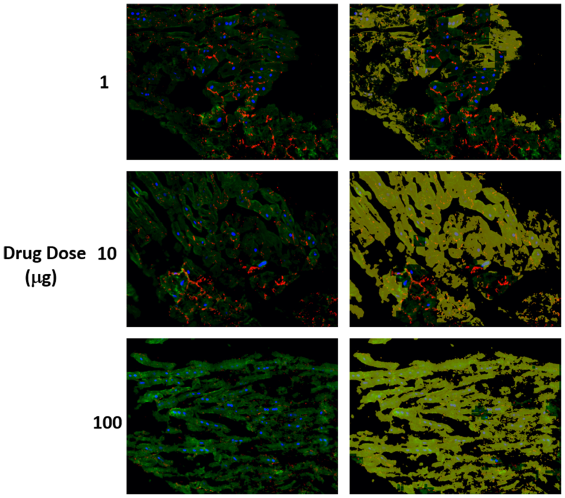
FIGURE 7.

The detected structural deterioration in the images of cells treated by herceptin. (a) is the original image where nuclear DAPI (Blue), cardiac troponin-T (green), and gap junction protein (connexin 43) (red). (b) is the original image with structural deterioration areas overlaid with yellow color that has opacity of 60%.
