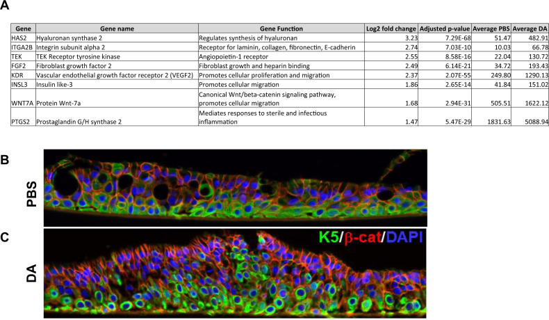
Fig. 4. DA vapor exposure induces an epithelial cell mobilization phenotype.
A Upregulated genes from the Positive regulation of epithelial cell migration gene ontology pathway that fell within the stringent cutoff of our dataset (FWER adjusted P value < 0.001, log2 fold change >1.45) B Representative confocal images of keratin 5 (K5, green, Alex Fluor 488), β-catenin (green, Alex Fluor 59) and nuclei (blue, DAPI) expression in NHBECs via immunofluorescent staining after PBS vehicle or C DA vapor exposure.

