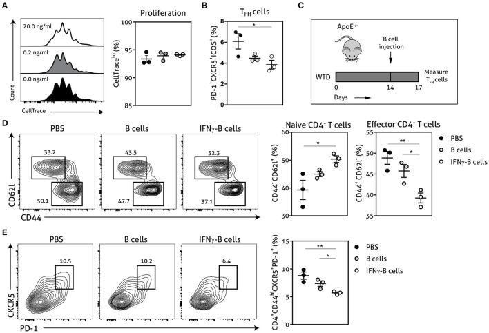
Figure 2.
IFNγ-B cells inhibit T follicular helper cells in vitro and in vivo. Isolated CD19+ B cells were unstimulated or stimulated with 0.2 ng/ml or 20.0 ng/ml IFNγ for 24 h and co-cultured with isolated CD4+ T cells. The coculture was stimulated with anti-CD3 (5 μg/ml) for 72 h after which (A) proliferation of CD4+ T cells was analyzed with CellTrace Violet and (B) the number of follicular CD4+ T helper (TFH) cells were analyzed. (C) apoE−/− mice were fed a Western type diet for 2 weeks after which they either received PBS, freshly isolated CD19+ B cells (B cells) or CD19+ B cells stimulated with 20.0 ng/ml IFNγ for 24 h (IFNγ-B cells). After three days, mice were sacrificed and spleens were isolated for flow cytometry analysis for (D) naive (CD62l+CD44−) and effector (CD62l−CD44hi) CD4+ T cells and (E) T follicular helper cells (CD4+CD44hiCXCR5+PD-1+). Representative flow charts of the CD4+ T cell population are shown. Data are analyzed with a One-Way ANOVA and shown as mean ± SEM (*p < 0.05, **p < 0.01). n = 3/group.

