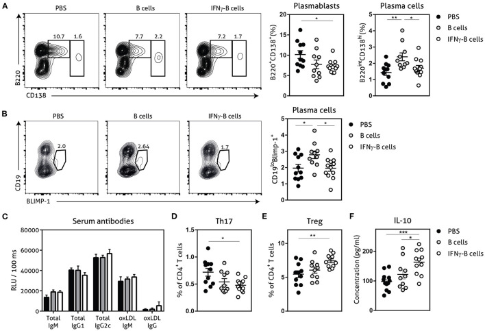
Figure 4.
Effects of adoptive transfer of IFNγ-B cells on the humoral immunity. ApoE−/− mice were fed a Western type diet for 7 weeks. After 2 weeks they received a perivascular collar and were treated with PBS, freshly isolated B cells (B cells) or B cells stimulated with 20.0 ng/ml IFNγ for 24 h (IFNγ-B cells). Mice received a total of three injections and injections were spaced every two weeks. After 7 weeks, mice were sacrificed and spleens were analyzed with flow cytometry. (A) Flow charts and quantification of plasmablasts (B220+CD138+) and plasma cells (B220loCD138hi). (B) Flow charts and quantification of CD19loBLIMP-1+ plasma cells. (C) Serum was analyzed for circulating antibodies by ELISA. Spleens were analyzed with flow cytometry for (D) Th17 cells (RORyt+) or (E) Treg cells (FoxP3+). (F) Splenocytes were isolated and stimulated with anti-CD3 (1 μg/ml) and anti-CD28 (0.5 μg/ml) for 72 h after which supernatant was collected and analyzed for cytokine expression with a multiplex analysis. Quantification of IL-10 concentration is shown. Data are analyzed with a One-Way ANOVA and shown as mean ± SEM (*p < 0.05, **p < 0.01). n = 10–12/group, ***p < 0.001.

