Fig. 6.

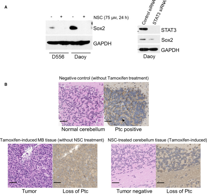
STAT3 activity is critical for Shh‐driven medulloblastoma formation in vivo. (A) Western blot of Sox2. Left panel: NSC74859 treatment abolished expression of Sox2 in both D556 and Daoy cells; right panel: STAT3 siRNA‐mediated knockdown of STAT3 led to decrease of Sox2 expression in Daoy cells. Western blot results shown are representative of at least three independent experiments. (B) Representative tissue histopathology from the Math1‐Cre‐ER‐Ptc floxed murine model of Shh MB. Left panels: H&E staining of normal cerebellum from Math1‐Cre‐ER‐Ptc floxed mouse without tumor (no tamoxifen induction) (top), cerebellar tumor after tamoxifen induction (without NSC74859 treatment) (bottom, left), and NSC74859‐treated tamoxifen‐induced mouse without cerebellar tumor (bottom right panels, left); right panels: immunohistochemistry for Ptc in cerebellar and tumor tissue of corresponding mice (brown staining, arrow in upper right panel points to examples of diffuse positive cell membrane expression in the external granular layer of normal cerebellum). Pictures shown are representative images; scale bars represent 50 µm.
