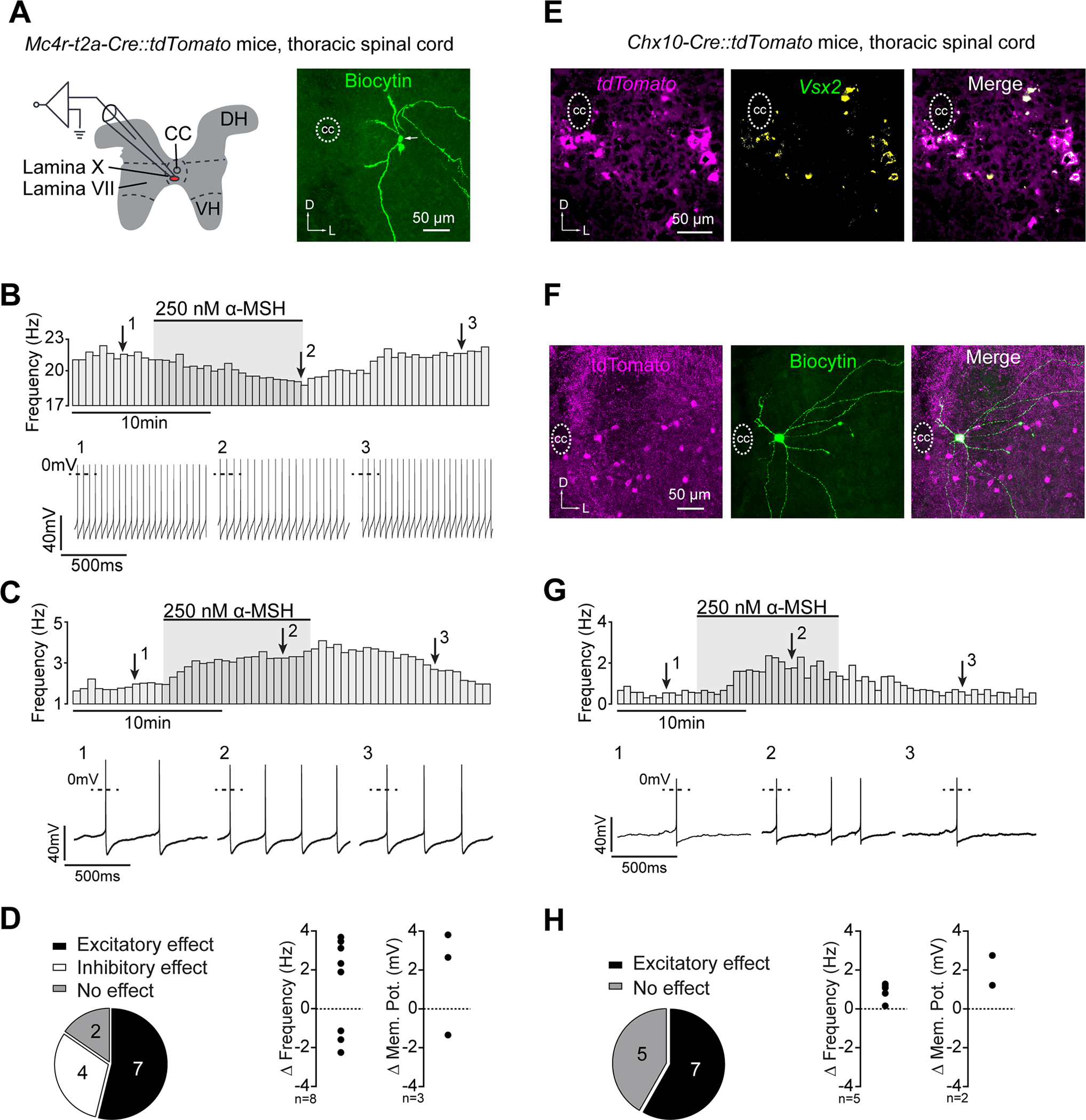
Figure 5: Mouse Mc4r and V2a neurons in spinal lamina X respond to α-MSH.

(A) Schematic illustration of the electrophysiological recordings from lamina X Mc4r::tdTomato neurons (left) and Biocytin-streptavidin staining of a recorded Mc4r::tdTomato neuron (right). (B,C) Rate histograms with the respective original recordings showing the inhibitory (B) and excitatory (C) effects of α-MSH on action potential frequency of lamina X Mc4r::tdTomato neurons. (D) Distribution of excitatory, inhibitory, and no responses (left) and relative effects of α-MSH on action potential frequencies or membrane potentials of spontaneously active or silent Mc4r::tdTomato neurons (right). (E) V2a::tdTomato neurons in Chx10-Cre::tdTomato mice as assessed by Vsx2 in situ hybridization. (F) Biocytin-streptavidin staining of a recorded V2a::tdTomato neuron. (G) Rate histogram with the respective original recording showing the excitatory effect of α-MSH on a V2a::tdTomato neuron. (H) Distribution of excitatory responses (left) and relative effects of α-MSH on action potential frequencies or membrane potentials of spontaneously active or silent V2a::tdTomato neurons (right).
The α-MSH (250 nM) effect was measured 8–10 minutes after starting the bath application. Neurons were classified as responders when the change in action potential frequency or membrane potential was 3x larger than standard deviation. CC, central canal; DH, dorsal horn; VH, ventral horn.
