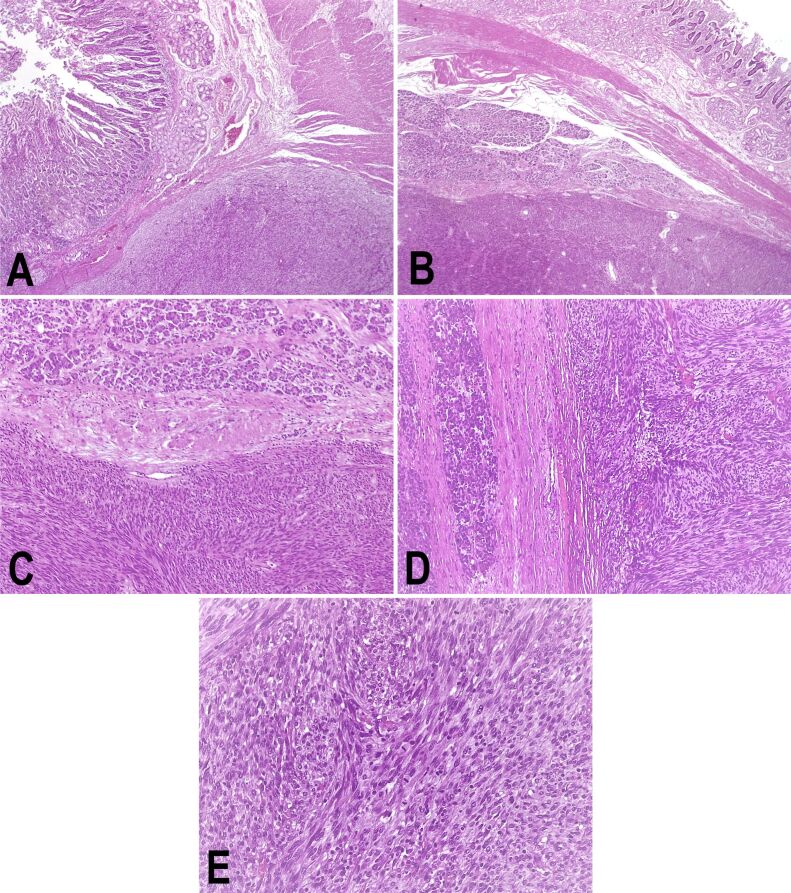
Figure 3.
Pancreatic GIST infiltrating the descending duodenum: (A) Duodenal wall and tumor; (B) Duodenal wall, pancreatic tissue, and tumor; (C) Pancreatic tissue (upper half) and tumor (lower half) (100x); (D) Pancreatic tissue and tumor proliferation; (E) Tumor proliferation, increased mitotic count. HE staining: (A and B) ×25; (C and D) ×100; (E) ×400. GIST: Gastrointestinal stromal tumor; HE: Hematoxylin–Eosin

