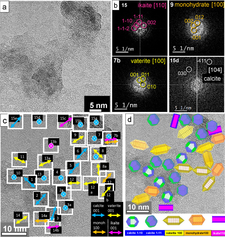
Figure 5.
Mineralized calcium carbonate polymorphs of an S-layer of G. stearothermophilus after 12 h incubation. (a) High-resolution TEM image of nanocrystals on the reassembled S-layer. (b) Selected-diffraction diagrams for kinetically grown samples with favored growth directions of ikaite [110], monohydrocalcite [100], vaterite [100], and an equilibrium orientation of calcite [104]. (c) Fourier analysis of the nanocrystals indicates different in-plane orientations. The crossed insets describe the [104], [214], [001], and [11̅2] axes of calcite (in blue), and the [001] axis of ikaite (in pink), both oriented parallel to the surface normal. (d) Model derived from panel c shows nanocrystal orientations of calcite (blue), vaterite (yellow), monohydrocalcite (orange), and ikaite (pink).

