Figure 9.
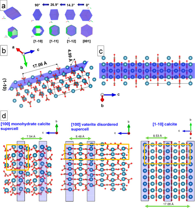
Calcite and precursor CaCO3 polymorphs growth on the S-layer. (a) Models of calcite nanocrystal orientations found in the image. Top: Rhombohedral calcite habit. Numbers between the arrows are indicating angles between the different orientations. Bottom: Calcite crystal habit with an additional set of prismatic facets {11̅0}. (b, c) (11̅0) surface of calcite is shown, which is in contact with the bacterial S-layer as well as with a vaterite particle. (d) Comparison of surfaces and summary of possible epitaxial relations between monohydrocalcite, vaterite, and calcite. Specific columns of Ca atoms along the b-axis are highlighted in blue. These Ca atoms are lying on the same upper level having a lateral distance of 7.54–8.52 Å. The Ca atoms between the columns (not highlighted) are positioned below this surface (2.5 Å or lower). Unit cells have been indicated in orange.

