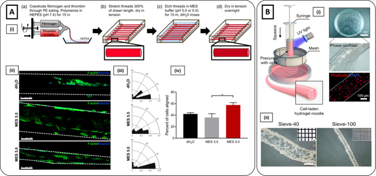
Figure 6.
Hydrogel extrusion methods. (Ai) Schematics of the fabrication of fibrin microthreads with MES etching (pH = 5.5 or 5.0). (ii) Immunostaining of phalloidin (green) and Hoechst (blue) showed higher myoblast elongation on fibrin microthreads treated with MES 5.0 compared to those treated with MES 5.5 and dH2O microthreads (control). Scale bar 100 μm. (iii) Nuclear orientation and (iv) the percent of total cells aligned along the microthreads direction (0°–15°) demonstrated higher preferential orientation along the long axis for MES 5.0 treated microthreads (Reproduced with permission from ref (165). Copyright 2020 WILEY-VCH Verlag GmbH & Co. KGaA, Weinheim). (Bi) Schematics of the fabrication of hydrogel fibers inspired by the production process of traditional Chinese noodles. Phase-contrast and fluorescent images (phalloidin red, nuclei blue) showed cells successfully encapsulated into hydrogel microfibers. (ii) Brightfield microscopy images of microfibers obtained using a sieve with different pore sizes (300 μm left, 100 μm right) (Reproduced with permission from ref (169). Copyright 2019 The Authors, Springer Nature CCBY-NC-ND 4.0).

