Figure 8.
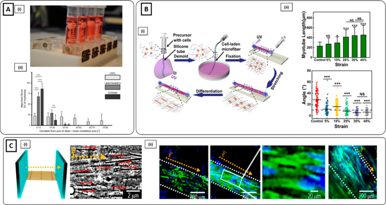
Mechanical and electrical stimulation on hydrogel-based fibers for SMTE. (Ai) C2C12-laden ring-shaped fibrin scaffold obtained by micromolding subjected to uniaxial mechanical stimulation through a spool–hook system working via magnetic force transmission. (iii) Assessment of myoblast alignment along the uniaxial direction revealed higher cell alignment for strained samples compared to unstrained (Reproduced with permission from ref (5). Copyright 2015 Elsevier). (Bi) Schematic representation of the cell-laden microfiber fabrication process and the uniaxial stretching of C2C12 myoblast laden microfibers. (ii) Cell-laden microfibers displayed an enhancement of myotube length and cell orientation when subjected to a strain higher than 35%. Scale bar 200 μm (Reproduced with permission from ref (72). Copyright 2019 American Chemical Society). (Ci) Schematics describing the direction of the electric field and the C2C12-laden GNWs/collagen scaffold and optical images showing the parallel distribution of GNWs. (ii) Immunostaining of MHC (green) and DAPI (blue) revealed alignment of myoblast along the electrical field direction (Reproduced with permission from ref (197). Copyright 2019 American Chemical Society).

