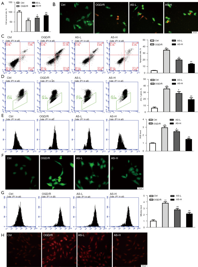
Figure 3.
AS protects against OGD/R-induced injury in HL-1 cardiomyocytes. (A) Cell survival rates were detected by the CCK-8 assay. (B) Fluorescence microscopy images of live/dead staining (green staining indicates viable cells, red staining indicates dead cells). (C) Cell apoptosis was evaluated using flow cytometry. (D) Depolarization of mitochondrial membrane potential was analyzed by flow cytometry. (E,F) The level of intracellular ROS was assessed using flow cytometry, representative images of ROS staining were captured under a fluorescent microscope. (G,H) The level of mitochondrial superoxide was determined using flow cytometry, representative images of MitoSOX staining were captured under a fluorescent microscope. Error bars indicate means ± SD of 3 independent experiments. **P<0.01, OGD/R group vs. control group; #P<0.05 or ##P<0.01, various dosages of AS pretreatment groups vs. OGD/R group. AS, asiaticoside; OGD/R, oxygen-glucose deprivation/reperfusion; CCK-8, Cell Counting Kit-8; ROS, reactive oxygen species; SD, standard deviation; L, low-dose; H, high-dose.

