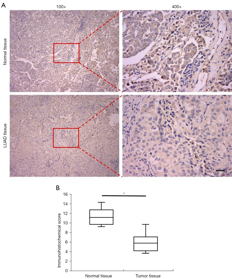
Figure 1.
IHC detection of REV-ERBα in normal tissue and tumor tissue in patients with lung adenocarcinoma. (A) Representative IHC staining images of REV-ERBα in normal tissue and tumor tissue at the magnification of ×100 and ×400 respectively (scale bar, 50 µm). (B) Statistics of IHC scores of expression of REV-ERBα in lung cancer tissue and paired non-cancerous tissues (n=66). (*P<0.05 compared with paired non-cancerous tissues). The slids were stained with 3,3’-diaminobenzidine (DAB) and counterstained with hematoxylin dye. IHC, immunohistochemical.

