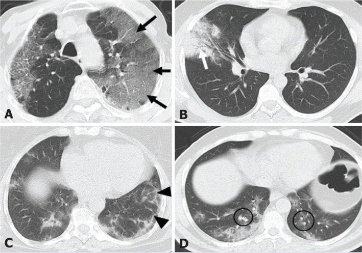
Figure 1.

The axial section of the chest computed tomography scan shows typical findings (A) ground glass opacity, crazy paving pattern (black arrows); (B) consolidation, air bronchogram (white arrow); (C) reticular pattern (black arrow-heads); (D) pulmonary vascular enlargement (black circle).
