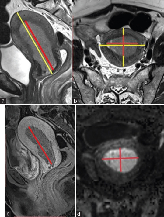
Figure 2.

(a and b) 63-year-old female with Grade III endometrioid carcinoma: Axial oblique T2-weighted image images and T2 sagittal images used to assess uterine volume (yellow line) and tumor volume (red line) (c-d) 63-year-old female with Grade III endometrioid carcinoma: Postcontrast 3 min delayed sagittal image and Axial oblique high B value (b = 1400/mm2) value diffusion images used for best tumor and myometrial contrast and measure tumor volume (red lines)
