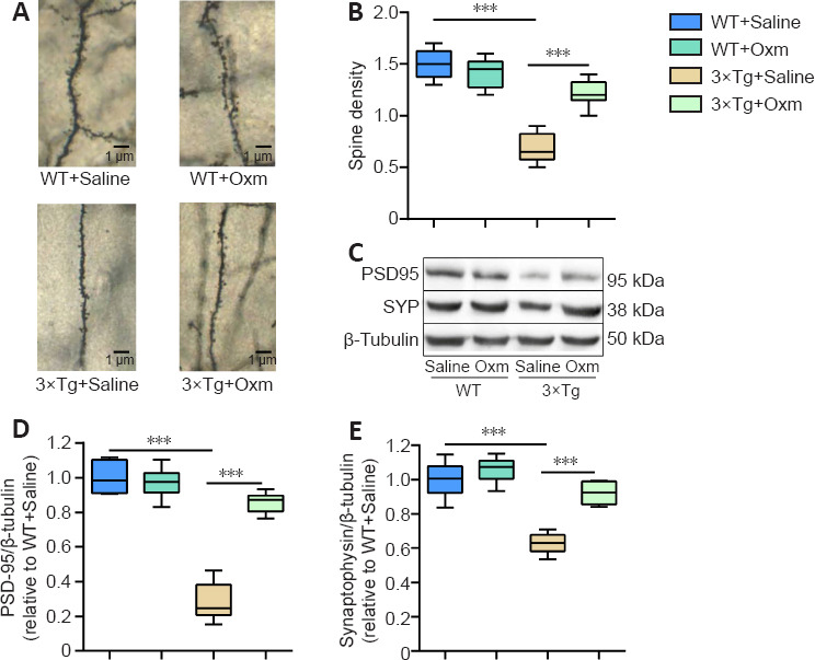
Figure 5.

(D-Ser2) Oxm increases the density of dendritic spines and the level of synaptic proteins in the hippocampal CA1 region of 9-month-old 3xTg mice.
(A) Representative images of dendritic spines in the hippocampal CA1 region of mice. Dendritic spine density was increased in the 3xTg + Oxm group compared with the 3xTg + saline group. Black arrows indicate representative dendritic spines. Scale bars: 1 μm. (B) Dendritic spine density in the hippocampal CA1 area (n = 4). (C) Representative bands showing the expression levels of PSD-95 and SYP in the hippocampus. β-Tubulin was used as a loading control. (D, E) Quantitative analysis of PSD-95 and SYP protein expression (n = 6). All values are shown as mean ± SEM, ***P < 0.001. Statistical comparisons were performed using two-way analysis of variance and post hoc Tukey's multiple comparison tests. 3xTg: APP/PS1/tau; Oxm: oxyntomodulin; PSD-95: postsynaptic density protein 95; SYP: synaptophysin; WT: wild-type.
