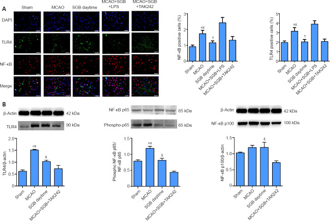
Figure 4.
Immunofluorescence and western blot analysis results reflecting the effect of SGB in ischemic stroke diabetic rats.
(A) Representative images of double immunofluorescence staining of TLR4- (FITC, green) and NF-κB-positive cells (Cy3, red) and DAPI for nucleus staining (blue). The NF-κB is expressed in both the nucleus and the cytoplasm, and the red color is seen in the nucleus, overlapping with the DAPI stained nucleus. Numbers of NF-κB- and TLR4-positive cells were higher in the MCAO group than in the SGB daytime group. Scale bars: 50 µm. *P < 0.05, vs. Sham group; #P < 0.05, vs. SGB daytime group; †P < 0.05, vs. MCAO + SGB + LPS group (one-way analysis of variance followed by the Student-Newman-Keuls post hoc test). (B) Quantitative analysis of western blots. The data are presented as the mean ± SD (n = 6). *P < 0.05, vs. Sham group; #P < 0.05, vs. SGB daytime group; †P < 0.05, vs. MCAO + SGB + TAK242 group (one-way analysis of variance followed by the Student-Newman-Keuls post hoc test). DAPI: 4′,6-Diamidino-2-phenylindole; FITC: fluorescein isothiocyanate isomer; LPS: Lipopolysaccharide, a TLR4 agonist; MCAO: middle cerebral artery occlusion; NF-κB: nuclear factor kappa-B; SGB: stellate ganglion block; TAK-242 (Resatorvid): a small-molecule-specific inhibitor of TLR4 signaling; TLR4: Toll-like receptor 4.

