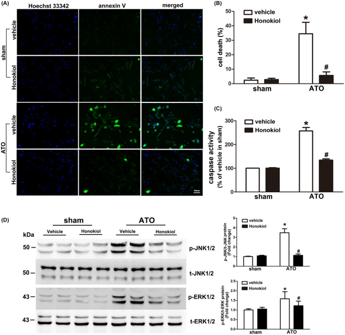
FIGURE 2.

Effects of Honokiol on ATO‐induced apoptosis in primary cultured cardiomyocytes. Cardiomyocytes were pretreated with Honokiol for 6 h followed by sham or 4 μM ATO treatment, inhibition of ATO‐induced apoptosis by ATO was assessed through Hoechst 33342(blue)/annexin V (green) staining. Cell images were taken under a fluorescence and UV light microscope, blue spots represent cell nuclei and green spots represent apoptotic bodies (A); the rate of cell death was counted under microscope (B); activity of caspase‐3 enzyme was measured using a fluorometric assay (C). The expression levels of phosphorylated and total ERK1/2 and JNK1/2 were detected by western blot analysis. A representative western blot was shown, quantitative data of p‐JNK/T‐JNK (upper) and p‐ERK/T‐ERK (lower) ratio were shown at right side. Lysates from primary cultured cardiomyocytes was collected 24 h after treatment from each group. Honokiol pretreatment significantly attenuated ATO‐induced phosphorylation of JNK1/2 and slightly inhibited phosphorylation of ERK1/2, collectively (D). The data are expressed as means ± SD from five independent experiments. *p < .05 versus vehicle in sham; # p < .05 versus vehicle in ATO‐treated cells
