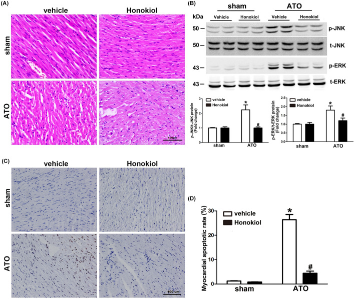
FIGURE 5.

Effect of Honokiol on myocardial apoptosis in mice exposed to ATO. Myocardial tissue sections of left ventricular specimens stained with HE (A), the characteristics of ATO cardiotoxicity were found in left ventricular sections, while HKL pretreatment abrogated the pathological changes made by ATO. Heart lysates were analyzed by western blotting for the phosphorylated and total ERK1/2 JNK1/2, Myocardial ERK1/2, and JNK1/2 phosphorylation level were markedly increased by 4 weeks of ATO exposure, which was inhibited by HKL pretreatment (B). Myocardial tissue sections of left ventricular specimens stained with TUNEL (C), heart sections from ATO group exhibited higher percentage of apoptotic cells. Pretreatment with HKL significantly decreased the percentage of apoptotic myocardial cells (Magnification = 200×). Apoptosis index was counted by two independent pathologists (D). The data are expressed as means ± SD, n = 6–8, *p < .05 versus vehicle in sham; # p < .05 versus vehicle in ATO‐exposed mice
