
FIGURE 6.
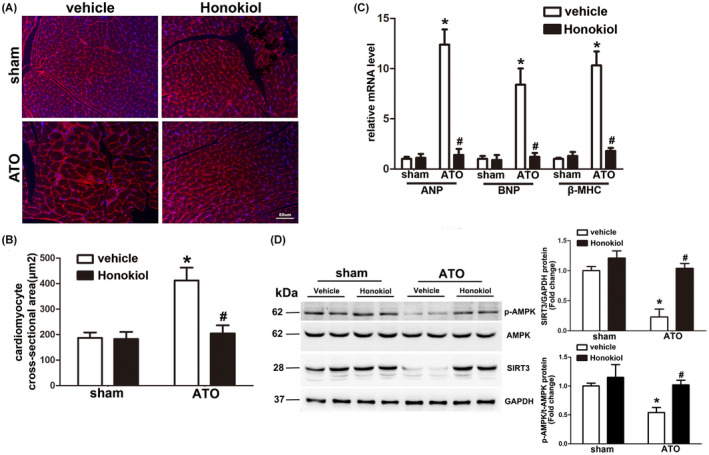
Honokiol pretreatment attenuated cardiac hypertrophy induced by arsenic trioxide exposure. Representative images of heart sections stained with WGA (red) and Hoechst 33342(blue) showed that cell size of cardiomyocytes increased by 4 weeks of ATO exposure, which was obviously attenuated by Honokiol pretreatment (A). Statistical results for quantification of the cardiomyocyte size in cross‐sectional area (B). Quantification results for real‐time PCR analysis of hypertrophic marker genes including ANP, BNP, and β‐MHC from hearts of mice in the indicated groups (C). Heart lysates were analyzed by western blotting for the indicated antibodies. Myocardial AMPK phosphorylation level and the expressions of SIRT3 were markedly downregulated by 4 weeks of ATO exposure, which was reversed by Honokiol pretreatment (D). The data are expressed as means ± SD, n = 6–8, *p < .05 versus vehicle in sham; # p < .05 versus vehicle in ATO‐exposed mice