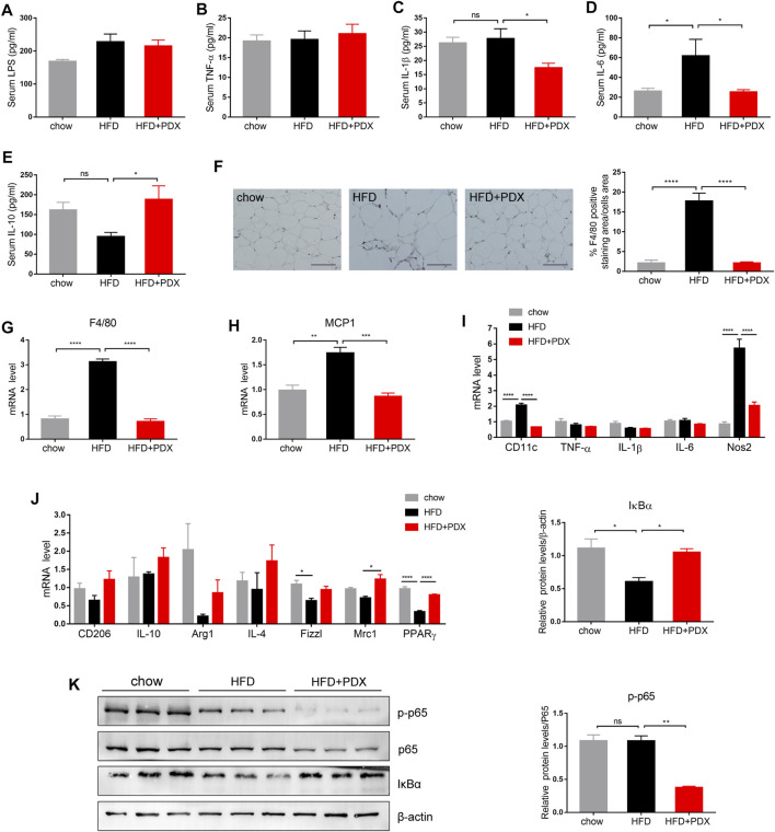
FIGURE 4.
PDX maintained a lower inflammation level and promoted M2 macrophage polarization in the adipose tissue of HFD-fed mice (Part 1). (A) Serum LPS. (B) Serum TNF-α (B). (C) Serum IL-1β. (D) Serum IL-6. (E) Serum IL-10. (F) Representative images of immunohistochemical staining of epididymal adipose tissue against the specific macrophage maker F4/80 and the quantification of F4/80-positive staining area. Scale bar, 100 μm; magnification, x200. (G) qPCR analysis of the mRNA levels of F4/80 in epididymal adipose tissue. (H) qPCR analysis of the mRNA levels of MCP1 in epididymal adipose tissue. (I) qPCR analysis of the mRNA levels of M1 macrophage maker CD11c, TNF-α, IL-1β, IL-6 and Nos2 in epididymal adipose tissue. (J) qPCR analysis of the mRNA levels of M2 macrophage maker CD206, IL-10, Arg1, IL-4, Fizz1, Mrc1 and PPARγ in epididymal adipose tissue. (K) Western blot analysis of the proteins expression involved in NF-κB signaling pathway in epididymal adipose tissue. n = 12 (A–F), n = 4 (G–J) or n = 3 (K) per group. * p < 0.05; ** p < 0.01; *** p < 0.001; **** p < 0.0001; ns, not statistically significant.

