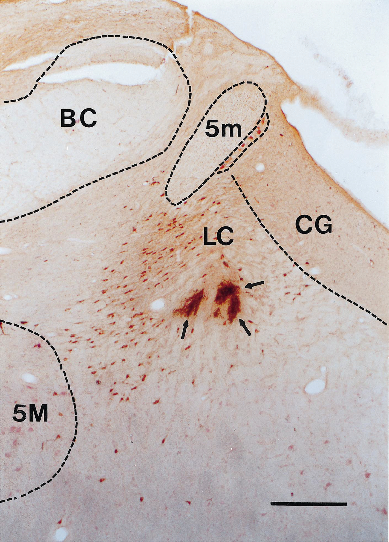
Fig. 1.

Coronal section at R3 level showing lesion marks made at the electrode tips (arrows) in a region of TH-positive cells (stained brown). Scale bar = 0.5 mm. BC, brachium conjunctivum; CG, central gray; 5M, motor trigeminal nucleus; 5m, tract of the mesencephalic trigeminal nucleus.
