
FIG. 6.
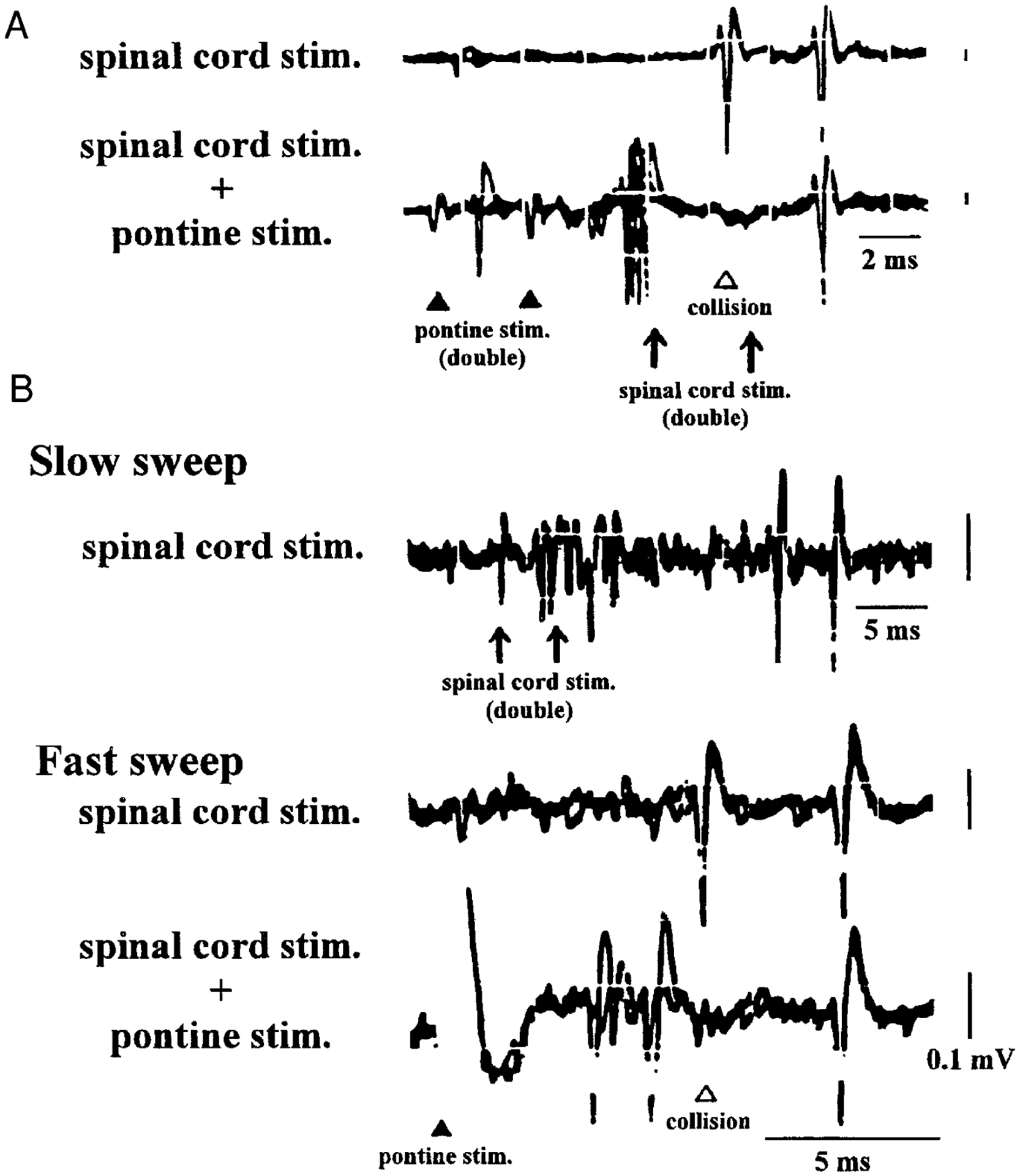
Action potentials of representative fast (A) and slow (B) conducting reticulospinal units are shown. A, top trace: action potentials activated antidromically by double spinal cord stimulation. Constant latency and the ability to follow high-frequency stimulation (330 Hz) are verified. On the bottom trace, the 1st action potentials antidromically activated by the 1st stimulation of the double spinal cord stimulation collide with action potentials orthodromically activated by double pontine stimulation. The 2nd action potential of the unit antidromically activated by the 2nd stimulation of the double spinal cord stimulation can still be identified. B: top trace shows the antidromic activation of the unit by double spinal cord stimulation (250 Hz) with a slow sweep. The 2nd and the 3rd traces present the same unit with a faster sweep with the same double spinal cord stimulation. In the 2nd trace, the constant latency and the ability to follow high-frequency stimulation can be seen. In the 3rd trace, the pontine stimulation is combined with double spinal cord stimulation. Pontine-induced spikes collide with antidromic spikes that are activated by the 1st spinal cord stimulation. Arrows, spinal cord stimulation at the L1 level; closed arrowheads, pontine stimulation; open arrowhead, pontine stimulation. Calibration: 2 (A) and 5 ms (B), 0.1 mV.