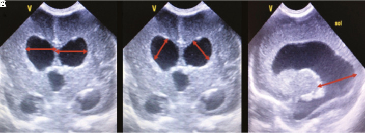
Figure 1.a-c.
Cranial ultrasound scans in a preterm infant with post-hemorrhagic ventricular dilatation. Figures represent the measurement points. (A) Ventricular index is the horizontal distance between the outermost part of the lateral ventricle and the interhemispheric fissure on the coronal scan at the level of the foramen of Monro. (B) Anterior horn width is the longest diagonal distance between the frontal horns of the lateral ventricles on the coronal scan at the level of the foramen of Monro. (C) Thalamo-occipital distance is the measurement between the most posterior portion of the thalamus and the occipital horn of the lateral ventricle in the parasagittal view.

 Content of this journal is licensed under a