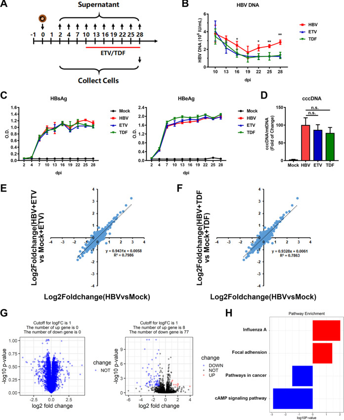
FIG 5.
Late administration of nucleot(s)ide analogues had limited effects on relieving HBV cytotoxicity. (A) 5C-PHH cells were infected with HBV (MOI of 200) and then treated with or without ETV (1 μM) or TDF (5 μM) at 10 dpi. (B and C) Supernatants were collected every 3 days and applied to HBV DNA (B) and HBsAg and HBeAg (C) analysis. (D) Intracellular cccDNA was extracted and detected by qPCR with indicated primers. (E) Comparison of ETV-treated or untreated HBV-infected cells with mock-infected cells for 28 days. Values were log2 transformed. (F) Comparison of TDF-treated or untreated HBV-infected cells with relative mock-infected cells. (G) Volcano plots displaying pairwise comparisons between early ETV-treated and untreated HBV-infected cells (left, 7 dpi; right, 28 dpi). (H) Gene set enrichment analysis (GSEA) pathway terms enriched among genes rescued by ETV therapy at 28 dpi are displayed.

