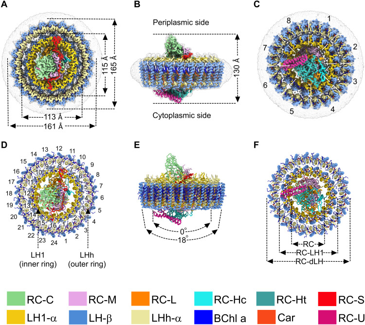
Fig. 1. Cryo-EM structure of the RC-dLH complex from G. phototrophica.
(A to C) Views of the color-coded RC-dLH density map. Detergent and other disordered molecules are in gray. (A) Perpendicular view from the periplasmic side of the membrane, with the diameters of the two LH rings indicated. (B) View within the plane of the membrane showing the overall height of the complex. (C) Perpendicular view from the cytoplasmic side. White dashed lines indicate pseudo-C8 symmetry of the double LHh-LH1 ring. (D to F) Ribbon models corresponding to (A) to (C). In (D), the LH1 and LHh subunits are numbered in black. (E) The two arrows indicate the degree of curvature of the outer and inner LH rings. (F) Concentric nature of the complex proceeding outward from the central RC. The color code is shown below the panels. Images were produced using ChimeraX (15).

