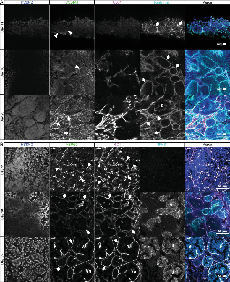
Figure 2. Sequential assembly of basement membrane components.
(A) Confocal immunofluorescence microscopy of wild-type kidney organoids showing the temporal emergence and co-distribution of COL4A1 and pan-laminin, and (B) perlecan and nidogen at days 11, 18, and 25 of differentiation. NPHS1 and CD31 were used as markers for podocyte and endothelial cells, respectively, in glomerular-like structures (g). Arrowheads indicate interrupted BM segments, large arrows indicate diffuse BM networks, and thin arrows indicate intracellular droplets of proteins.

