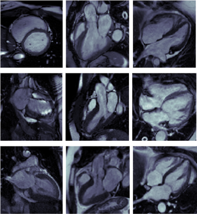
Figure 3:

Sample of misclassified cardiac MRI images.
Top: Images Misclassed by both DL and ML; Left: SAX image misclassed as 3-Ch by DL and 4-Ch by ML; Center: 3-Ch image misclassed as SAX by DL and 2-Ch by ML; Right: 4-Ch image misclassed as 3-Ch by both DL and ML
Middle: Images Misclassed by DL only; Left: 2-Ch image misclassed as SAX; Center: 3-Ch image misclassed as SAX; Right: 4-Ch image misclassed as 3-Ch
Bottom: Images Misclassed by ML only; Left: 2-Ch image misclassed as SAX; Center: 3-Ch image misclassed as SAX; Right: 4-Ch image misclassed as 3-Ch
