Figure 3.

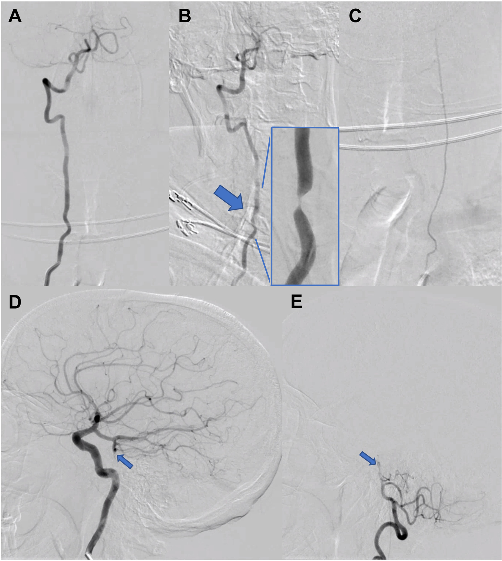
A-B. Digital subtraction angiography revealed the right vertebral artery was unremarkable in the standard AP position (A) but turning the head to the right elicited a focal region of stenosis (B, large arrow, magnified view is inset). C. The left vertebral artery was congenitally hypoplastic. AP view with left vertebral artery injection. D-E. There was a chronic-appearing mid-basilar occlusion (small arrows), and the bilateral posterior cerebral, bilateral superior cerebellar, and superior basilar arteries were supplied by the anterior circulation through a robust right posterior communicating artery. Both are lateral views; D is right internal carotid artery injection; E is right vertebral artery injection.
