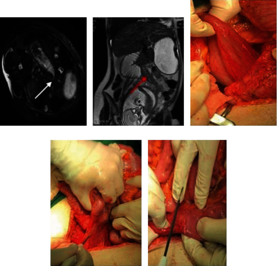
Figure 1.

(a) Transverse oblique (patient is lying on a side) view marked dilation of the stomach and duodenum up to D3 level (the arrow is the point of stenosis). (b) The site of Ladd's bands (red arrow), uterus, and duodenum. (c) Dilated duodenum under cecum. (d) Peritoneal Ladd's bands. (e) Released peritoneal band in the site of stenosis.
