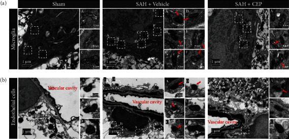
Figure 6.

The mitochondrial damage in microglia and endothelial cells after SAH and the rescue effects of CEP. (a, b) Representative TEM picture of mitochondria in microglia and endothelial cells. Microglia presented irregular nuclear morphology (bean or jagged-shaped), electron-dense cytoplasm (dark cytoplasm), and distinct heterochromatin pattern (thick and dark chromatin condensation beneath the nuclear membrane). Endothelial cells were determined by typical vascular structure and its internal residual blood components. The red arrow points to the shrunken mitochondria with increased mitochondrial membrane density, vanishing cristae, and collapse of outer membrane. The white dashed frame marked with capital letters in the left overall image (scale bar = 1 μm) corresponds to the scattered images (scale bar = 0.2 μm) enlarged on the right.
