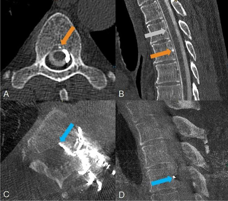
Fig. 1.
A 60-year-old man with a central CVF at the level of Th5/6 left. A, B Left lateral decubitus CTM shows a hyperdense spot in the axial and sagittal reformatted images (orange arrow) at the level of Th 5/6 representing the proximal “hyperdense basivertebral vein” sign. B, One level above e.g. the triangular shape of the basivertebral vein is empty (grey arrow). C, D After transvenous embolization the Onyx cast is confirmed to be within the central draining CVF in axial and sagittal reformatted images of a cone beam CT in which it fills the proximal basivertebral vein (blue arrow) brightly

