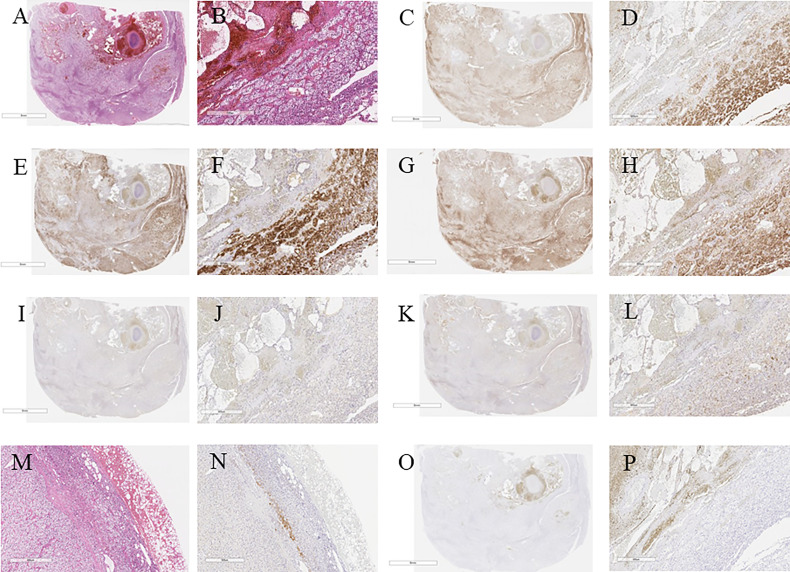
Figure 3.
(A) Hematoxylin and eosin immunostaning. (B) Hematoxylin and eosin-stained tumor section on high magnification showing cortical adenoma (40×). (C) c17 immunostaining. (D) c17-stained tumor section on high magnification (40×). (E) HSD3B2 immunostaining. (F) HSDB2-stained tumor section on high magnification (40×). (G) CYP11B1 immunostaining. (H) CYP11B1-stained tumor section on high magnification (40×). (I) HSDB1 immunostaining. (J) HSDB1-stained tumor section on high magnification (40×). (K) DHEA-ST immunostaining. (L) DHEA-ST-stained tumor section on high magnification (40×). (M) Hematoxylin and eosin-stained concomitant adrenal tissue on high magnification (40×). (N) High magnification of DHEA-ST-stained concomitant adrenal tissue on high magnification (40×). (O) CYP11B2 immunostaining. (P) CYP11B2-stained tumor section on high magnification (40×).

