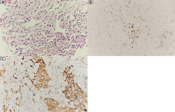
Figure 1.
Histological and immunohistochemical examination of the specimen from the right lymph lesion. (A) A representative image of the tumor specimen with H&E staining. The original tissue specimen and an H&E staining image with ×40 original magnification are provided in Supplementary Figure S2. (B, C) Representative images of immunohistochemical staining for (B) TTF-1 and (C) CK7 which demonstrate positive results. All images are ×400 original magnification. The immunohistochemical results indicate a great probability of metastatic adenocarcinoma for the lymph lesion.

