
Figure 4.
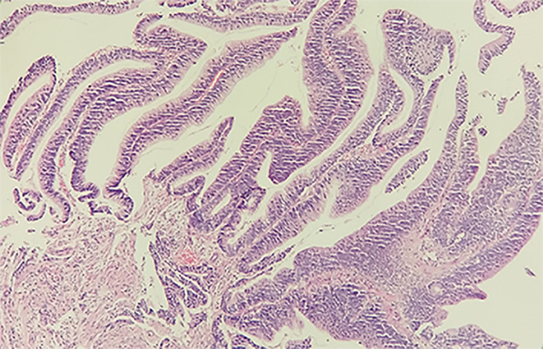
Histological examination of the specimen from the rectosigmoid junction with H&E staining. The tissue is 0.3 × 0.3 × 0.2 cm3 in size, grayish-white in color and tough in texture. The high-grade tubulovillous adenoma in the rectosigmoid junction supports the PET/CT diagnosis.