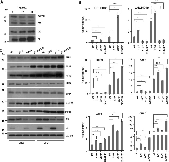
Fig. 2. Loss of CHCHD2 and CHCHD10 promotes mitochondrial integrated stress response.
A Whole-cell lysates of HeLa cells treated with DMSO or CCCP (10 mM, 12 h, or 24 h) respectively were analyzed for indicated protein expression by immunoblot. GAPDH was used as a loading control. B HeLa cells were infected with control, shCHCHD2, shCHCHD10, shCHCHD2/CHCHD10 lentiviral particles respectively and cultured for 7 days, treated with CCCP (10 mM) for 24 h, and then relative transcript levels of the indicated gene was analyzed by RT-QPCR. Statistical significance analysis was used by two-way ANOVA with Sidak’s multiple comparisons test, *P < 0.05, **P < 0.01, ***P < 0.001, N.S. not significant. C HeLa cells were infected with control, shCHCHD2, shCHCHD10, shCHCHD2/CHCHD10 lentiviral particles respectively and cultured for 7 days, treated with CCCP (10 mM) for 24 h, and then the indicated protein expression was analyzed by immunoblot. GAPDH was used as a loading control.

