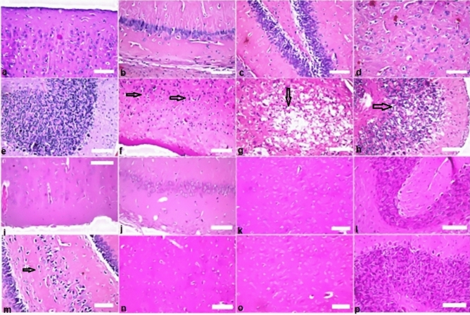
Figure 9.
Histopathological findings in brain (a–e) showing normal histopathological of the cerebral cortex of rat. (a) The control group showing normal appearance of the nerve cells. (f–h) Rats which exposed to gamma rays at dose 6 Gy revealed nuclear pyknosis (arrows) (score 3), degeneration (score 2), and cellular vacuolization (score 2) were detected in the neurons of central cortex, striatum and cerebellum respectively. (i,j) Rats were administered BV 0.05 mg/kg showed no alteration (score 0) of the brain. (k,l) The group of animals were administered propolis (300 mg/kg): there was no histopathological alteration as recorded in brain. (m) Nuclear pyknosis and degeneration (arrow) (score 1) were in some neurons of fascia dentata in the group of rats administered propolis 300 + BV 0.05 mg/kg. (n–p) Normal histological structures (score 0) of brain occurred in the normal rats administered propolis, BV, and combined treatment (Pr + BV) respectively.

