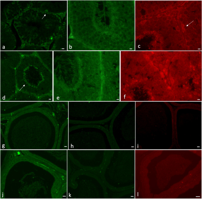
Figure 3.
Immunolocalization of docosahexaenoic acid (DHA), eicosapentaenoic acid (EPA) and arachidonic acid (ARA) in testis and cauda epididymis of rabbits fed control and FLAX diets. In (a) a clear DHA localization was in interstitial tissue, Sertoli cells and spermatogonia; after FLAX diet (d), the signal was also evident in elongated spermatids (arrow); in (b) the EPA label appeared localized in the germ cells at different stages of maturation; after FLAX diet, the label was intense in the same cells (e); in (c) the ARA signal was detected in interstitial cells, Sertoli cells (arrow) and elongated spermatids of control testis. The same labelling was present in testis from rabbits fed FLAX diet (f). In the epididymis of rabbits fed control (g,h,i) and FLAX diets (j,k,l), in (g,i,j and l), a limited number of vesicles in the lumen were labelled (DHA, g and j; ARA, i and l); on the contrary, in (h,k) a number of vesicles appeared labelled in the lumen where the spermatozoa were located (EPA). Bar: a–f, 10 µm, g–l, 50 µm.

