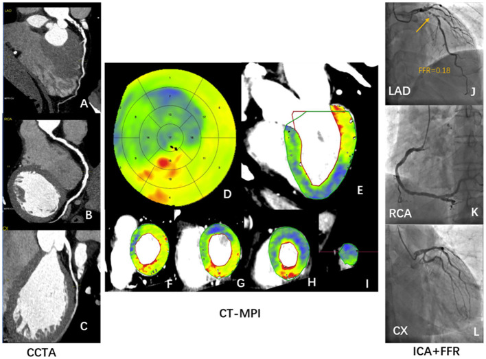
Figure 6.
Case illustrating hyperemic MBF can identify ischemic stenosis confirmed by ICA/FFR. A 56-year-old man who presented with a history of hypertension, current smoking, symptomatic for suspected angina, and a recent inconclusive 24 h' DCG. (A–C) Rest CCTA shows severe stenosis of distal LAD (A) and multiple mild stenosis of RCA (B). (D–I) Dynamic stress CT-MPI bull's eye diagram (D), long axis view (E), and short axis view (F–I) all show severe induced perfusion defects in the anterior wall, septum, and apical wall of left ventricle. The regional hyperemic MBF of LAD, RCA, LCX are 77 ml/100 mml/min, 126 ml/100 ml/min, and 107 ml/100 ml/min, respectively. (J–L) ICA shows severe proximal LAD stenosis (J) with positive invasive FFR (J). ICA shows multiple mild stenosis in RCA (K) and no stenosis of LCX (L). CCTA, coronary computed tomography angiography; CT-MPI, computed tomography myocardial perfusion imaging; DCG, dynamic cardiogram; FFR, fractional flow reserve; ICA, invasive coronary angiography; LAD, left anterior descending coronary artery; LCX, left circumflex coronary artery; MBF, myocardial blood flow; RCA, right coronary artery.

