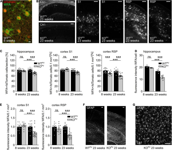
FIGURE 6.
Perineuronal networks (PNN) decrease in KCC2 KOPV mice. (A) Representative WFA stainings of the somatosensory cortex of a WTPV mouse at the age of 8 weeks. Scale bar 5 μm. (B) Representative WFA stainings of the hippocampus (left), the S1 (middle) and RSP cortex (right) of WTPV and KCC2 KOPV mice at 23 weeks of age. Scale bars 100 μm. (C) Quantification of WFA+/tdTomato cells in the hippocampus (left), S1 (middle), and RSP (right) cortex of WTPV and KCC2 KOPV mice at the age of 8 and 23 weeks shows a reduced proportion of ensheathed PV-INs in older KOPV mice (n = 15/15 slices, N = 5/5 mice; 1-way ANOVA; Bonferroni’s multiple comparison test; ns not significant; ***p < 0.001). (D,E) WFA signal intensity per individual cell in the hippocampus (D) (n = 12/9 slices; N = 4/3 mice; 1-way ANOVA; Bonferroni post-test; ns not significant; ***p < 0.0001). WFA signal intensity per 0.1 mm2 in the S1 cortex (left) and RSP (right) decreases over time in KCC2 KOPV mice (n = 13/15 slices; N = 5/5 mice; 1-way ANOVA Bonferroni post-test; ns not significant; ***p < 0.0001). (F) Representative GFAP staining of the cortex and hippocampus of WTPV and KCC2 KOPV at the age of 23 weeks. Scale bar 100 μm. (G) Close up of GFAP -stained reactive astrocytes in CA3 in KCC2 KOPV. Scale bar 50 μm.

