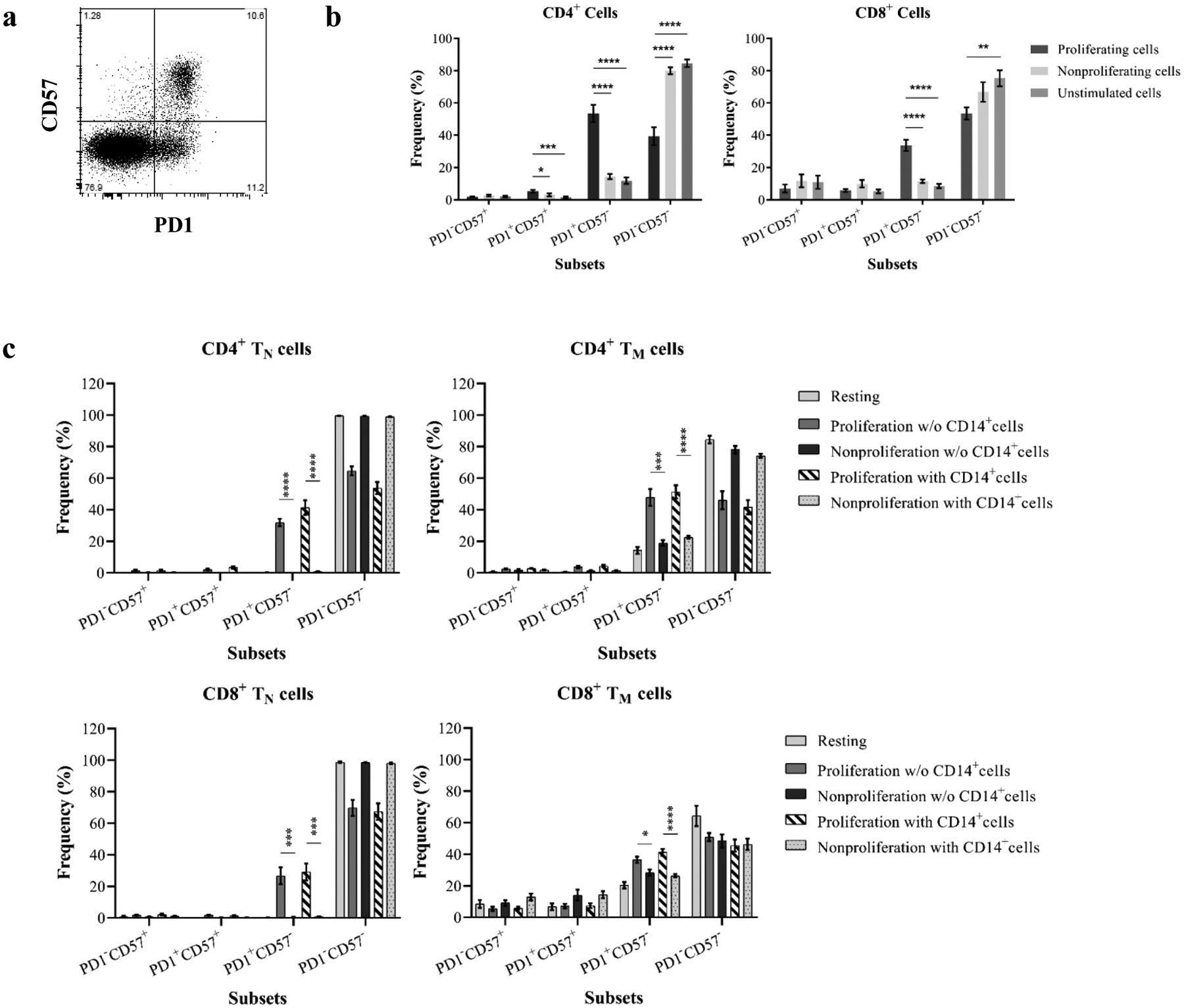
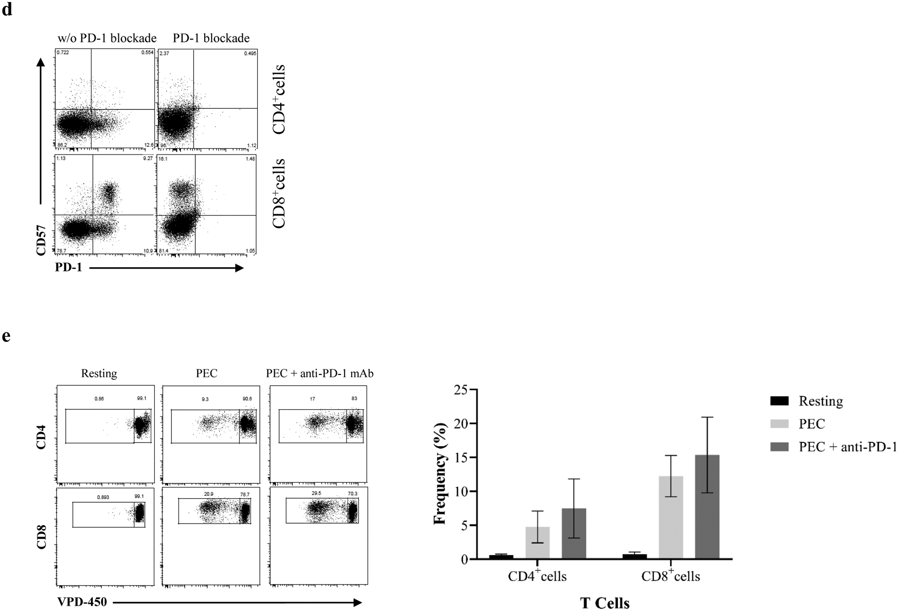
Figure 6. Upregulation of PD-1 expression on xenoreactive proliferating T cells.


(a) CD4+ and CD8+ cells are segregated into four subsets based on surface expression of PD-1 and CD57. (b) Both CD4+ and CD8+ cells significantly upregulate PD-1but not CD57 expression following stimulation by PECs. (c) Purified naïve and memory cells were analyzed for PD-1 expression following induction of proliferative responses by PECs. An increased PD-1 expression on proliferating cells was detected in CD4+ and CD8+ cells. (d) A representative sample shows that PD-1–specific antibody completely blocks PD-1 expression on CD4+ and CD8+ cells. (e) VPD-450–labeled responder PBMCs were stimulated with PEC in the absence or presence of PD-1 specific antibody for 6 days. A representative from one individual showed the xenoreactive T cell proliferation with or without PD-1 specific antibody (left). The blockade of PD-1 expression with PD-1 specific antibody is not effective to enhance xenoreactive T cell proliferation in response to PEC. (* p<0.05, ** p<0.001, *** p<0.0001, ****p<0.00001)
