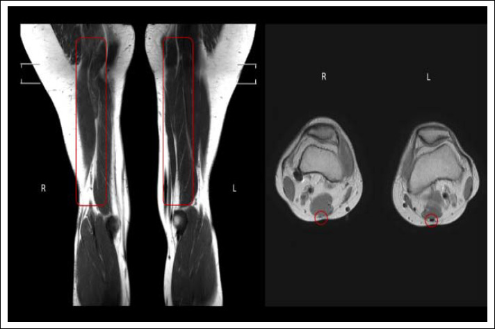
Figure 2.

On the right, coronal MRI of right (R) and left (L) limbs with highlighted semitendinosus muscle in one of the participants with ACL reconstruction of the right limb. On the left, transverse MRI with highlighted distal semitendinosus insertion. No regeneration was observed for the harvested tendon.
