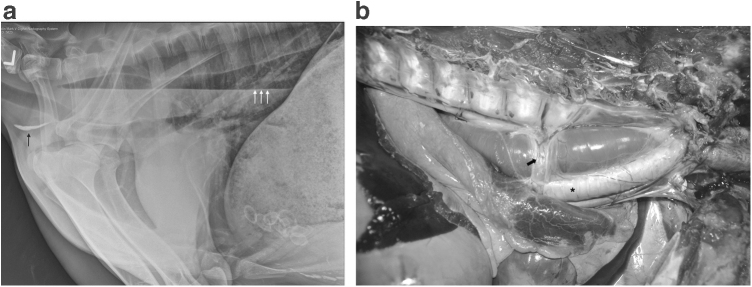
FIG. 1.
Megaesophagus. (a) Left lateral thoracic radiograph showing diffuse distention of the thoracic esophagus. The esophagus contains fluid and gas that creates a gas–liquid interface (white arrows) as well as gravity-dependent mineral debris (black arrow). (b) Gross necropsy image of the esophagus, located between the vertebral column dorsally (top of photo) and the trachea ventrally (asterisk), enlarged, and focally narrowed by the overlying azygos vein (arrow) where it courses over the esophagus.

