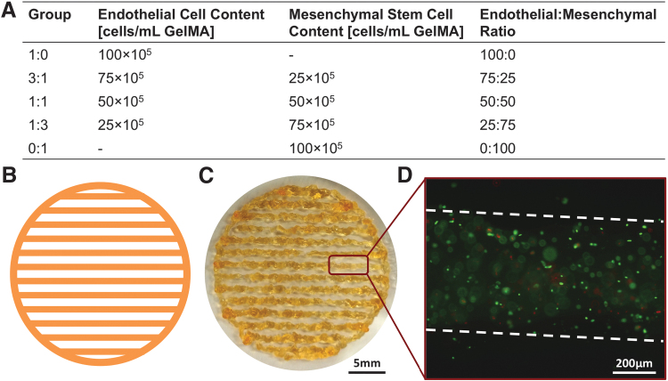
FIG. 1.
Experimental layout and scaffold fabrication. (A) EC and MSC were loaded in GelMA according to five different ratios, starting from a solution of PBS containing 7.5% w/v GelMA and an overall cell concentration of 106 cells/mL GelMA. (B) Scaffolds were first CAD designed and (C) subsequently fabricated using extrusion-based bioprinting. (D) Upon bioprinting, scaffolds were analyzed using Live/Dead staining to ascertain that the printing conditions did not negatively affect cell viability. In (D), live and dead cells are stained in green and red, respectively, while the broken white line indicates the fiber contours. EC, endothelial cell; GelMA, gelatin methacrylate; MSC, mesenchymal stem cell; PBS, phosphate buffered saline.

