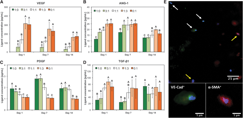
FIG. 4.
Expression of angiogenic/arteriogenic factors in bioprinted human cocultures. HUVECs and hMSCs were cocultured in five different ratios (1:0, 3:1, 1:1, 1:3, and 0:1) and analyzed after 1, 7, and 14 days for the expression of key angiogenic/arteriogenic factors using ELISA (A–D, respectively). In (A–D), data are reported as mean ± standard deviation (n = 4), and groups not connected by the same letter within each factor/time point tested are statistically different (p < 0.05). (E) Representative staining of bioprinted scaffold cultured at EC:MSC 3:1 ratio. Cells were stained for nuclei (blue), VE-Cad (green), and α-SMA (red). White and yellow arrows indicate HUVECs and hMSCs, respectively. High magnification insets show positive staining for VE-Cad in HUVECs (bottom left) and for α-SMA in hMSCs (bottom right). α-SMA, α-smooth muscle actin; ELISA, enzyme-linked immunosorbent assay; VE-Cad, vascular endothelial cadherin.

