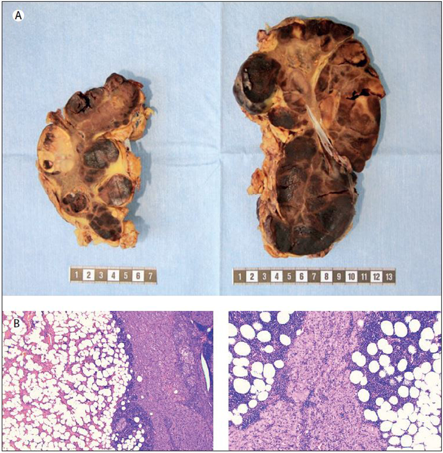
Figure 1.

Macroscopic and histological attributes of a bilateral adrenalectomy specimen in a patient with congenital adrenal hyperplasia. Top row: Gross image of the resected adrenals displaying multiple foci of adrenal myelolipoma (dark brownish-red areas) intermingled with hyperplastic adrenal cortical tissue (bright yellow). Metric ruler is depicted for size estimations. Bottom row: Representative photomicrographs of haematoxylin-eosin stained sections from each adrenal specimen (magnified x40 and x100 respectively) displaying myelolipoma and hyperplastic cortical tissue.
