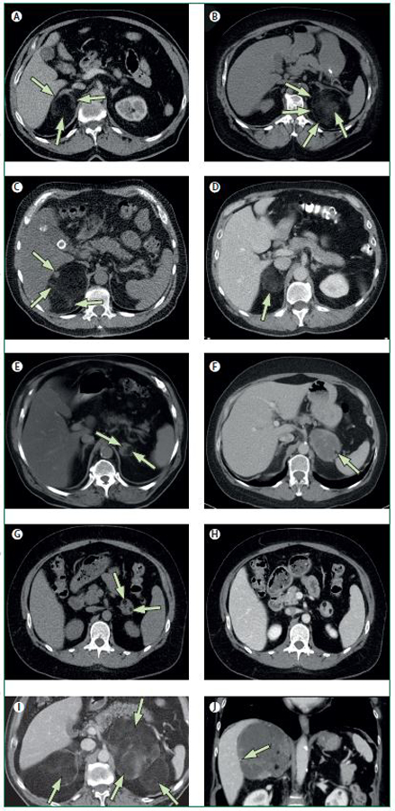
Figure 2.

A-I. Transversal computed tomography (CT) images. J. Coronal CT image.
A. Typical myelolipoma in the right adrenal gland (long arrow), comprising almost exclusively macroscopic fat, spreading the lateral and medial adrenal limbs apart (short arrows), extending posteriorly but demarcated from the periadrenal fat by a thin capsule.
B. The myelolipoma in the left adrenal gland is dislocating the lateral adrenal limb ventrally and extends laterally to the spleen. The capsule is clearly seen (short arrows), and the myeloid component shows a cloudy appearance (long arrow).
C. In the myelolipoma in the right adrenal gland, the myeloid components are seen as a nodule (long arrow) in the ventral-medial part of the tumour and as strands (small arrows) in its posterior aspects.
D. In this myelolipoma in the right adrenal gland (arrow), the myeloid and fat components are mixed and are evenly spread throughout the whole tumour.
E. By contrast, in this myelolipoma in the left adrenal gland, the myeloid (short arrow) and fat components (long arrow) are distinctly separated and are located in the medial and lateral part of the tumour, respectively.
F. The myelolipoma in the left adrenal gland comprise almost exclusively myeloid tissue in which there is a small island of macroscopic fat in the lateral-posterior aspect of the tumour (arrow).
G. CT without intravenous contrast-enhancement. In this myelolipoma in the left adrenal gland, with a similar appearance there are two small fatty islands (short arrows).
H. CT with intravenous contrast-enhancement in the venous phase shows in the same tumour contrast-enhancement of the myeloid component, similar to that of the liver and spleen.
I. Myelolipomas are sometimes bilateral, especially when they are large (arrows).
J. Sometimes calcifications are seen in myelolipomas and they are usually small (arrow).
