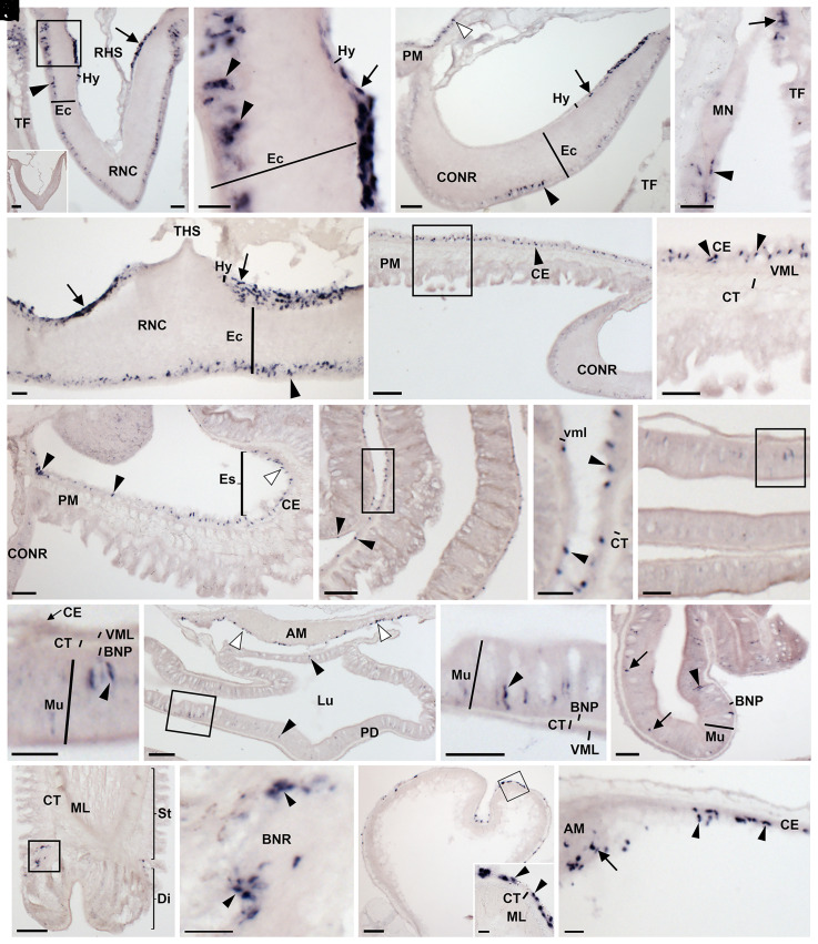
Fig. 4.
Localization of ArSSP1 mRNA in A. rubens using in situ hybridization. (A) Transverse section of radial nerve cord incubated with antisense probes showing ArSSP1-expressing cells in both the hyponeural region (arrow) and the ectoneural region (arrowhead). Stained cells can also be seen in an adjacent tube foot. The Inset shows that no staining is observed in a section incubated with sense probes, demonstrating the specificity of staining observed in sections incubated with antisense probes. (B) Higher-magnification image of the boxed region in A, showing stained cells in the hyponeural region (arrow) and in the subcuticular epithelial layer of the ectoneural region (arrowheads). (C) Transverse section of circumoral nerve ring showing stained cells in the hyponeural region (arrow) and the ectoneural region (arrowhead). Stained cells can also be observed in the coelomic epithelial lining of the peristomial membrane (white arrowhead). (D) Stained cells can be seen in the external epithelial layer of the marginal nerve (arrowhead) and in an adjacent tube foot (arrow). (E) Parasagittal longitudinal section of radial nerve cord showing stained cells in both the hyponeural region (arrows) and ectoneural region (arrowhead). (F) Transverse section of the central disk region showing ArSSP1-expressing cells in the coelomic epithelial lining of the peristomial membrane (arrowhead). (G) Higher-magnification image of the boxed region in F, showing stained cells in the coelomic epithelial lining of the peristomial membrane (arrowheads). (H) Transverse section of the central disk region showing stained cells in the esophagus (white arrowhead) and in the peristomial membrane (black arrowheads). (I) Transverse section of the central disk region showing stained cells in the cardiac stomach (arrowheads). (J) Higher-magnification image of the boxed region in I, showing round-shaped stained cells (arrowheads) adjacent to the visceral muscle layer. (K) Transverse section of the central disk region showing staining in the pyloric stomach. (L) Higher-magnification image of the boxed region in K, showing elongate-shaped stained cells in the mucosal layer (arrowhead). (M) Transverse section of the central disk region showing stained cells in the pyloric duct (black arrowheads) and in the coelomic epithelial lining of the apical muscle (white arrowheads). (N) Higher-magnification image of the boxed region in M, showing elongate-shaped stained cells in the mucosal layer (arrowhead). (O) Transverse section of an arm showing stained cells in a pyloric cecum. Elongate-shaped stained cells can be seen in the mucosal layer (arrowhead), and round-shaped stained cells (arrows) can be seen close to the position of the basiepithelial nerve plexus. (P) Longitudinal section of a tube foot showing stained cells at the junction between the stem and the disk region. (Q) Higher-magnification image of the boxed region in P, showing stained cells (arrowheads) are located around the basal nerve ring. (R) Transverse section of an ampulla of a tube foot showing stained cells in the coelomic epithelial lining. The Inset shows higher-magnification image of the boxed region, showing stained cells (arrowheads) in the coelomic epithelial lining of the ampulla. (S) Transverse section of the central disk region showing stained cells in the coelomic lining of the body wall (arrowheads) and the coelomic epithelial layer of the apical muscle (arrow). Abbreviations: AM, apical muscle; BNP, basiepithelial nerve plexus; BNR, basal nerve ring; CE, coelomic epithelium; CONR, circumoral nerve ring; CT, collagenous tissue; Di, disk; Ec, ectoneural region; Es; esophagus; Hy, hyponeural region; Lu, lumen; ML, muscle layer; MN, marginal nerve; Mu, mucosa; PD, pyloric duct; PM, peristomial membrane; RHS, radial hemal strand; RNC, radial nerve cord; St, stem. TF, tube foot; THS, transverse hemal strand; VML, visceral muscle layer. (Scale bars, 8 μm in R, Inset; 32 μm in A, B, D, E, J, and L; 60 μm in A, Inset, C, G, H, K, N, O, Q, R, and S; 120 μm in F, I, M, and P.)

